
Regular breast Self Examination is a very important exercise which every woman must add in her routine. While examining, even if there is the slightest doubt (check for signs and symptoms here), it must be investigated further. Immediate medical assistance should be sought, for correct information at the right time, means half the battle won already!
No single test can detect all breast cancers and hence it is prescribed to take the ‘triple-test’ at a hospital with trained medical professionals. The triple test, which might take a few hours, covers three major aspects of diagnosing the presence of cancer. Women nurses/doctor shall be with you when you get your tests done. You might want to have someone close accompany you during the clinical examination.
PRE-EXAMINATION STUDY
The specialist might gather the background data from you before starting the examination. This might include family history of cancer, symptoms you discovered, other health problems being faced, if you are under any medication among others.
THE TRIPLE TEST
Depending upon the case-requirement, the staff might want to perform one or more of the following tests to find out about the changes in your breast. Please note that you should not worry if you don’t have all the tests done on you, not everyone needs all the three tests.
1. CLINICAL BREAST EXAMINATION
 The first step is a clinical breast examination, where a doctor or nurse looks at and carefully feels your breasts and the area around them. You will be asked to take all your clothes off your top half. The clinician shall make you sit or lie down and shall feel your breasts all the way up to your collarbone and even armpits (where lymph nodes are present. Breast cancer can sometimes spread into lymph nodes). Any suspicion of cancer during clinical breast examination shall lead you to the next test.
2. MAMMOGRAM

‘Mammography‘ is a specific type of breast imaging that uses low-dose X-rays to detect cancer early. The result of the process is a mammogram, which is a picture of the inside of your breast. In general, regular mammograms aren’t recommended for women under 40 years of age, in part because breast tissue tends to be dense, making mammograms less effective. Here’s a quick explainer of the process:
- A female doctor/nurse shall be assisting you while using the machine.
- You will be asked to bare your top half, stand and face the mammogram machine.
- It has handles to hold on to, which keep your arms out of the way.
- The nurse shall place both your breasts on the bottom shelf of the machine and press it firmly with a clear plastic plate.
- The machine takes 2-3 pictures of each breast in this position.
- It is a slightly uncomfortable and painful process (for a few) but it’s normal and does not last long.
3. TISSUE DIAGNOSIS
Tissue diagnosis involves observing the involved breast tissue under a microscope. This is the most important investigation (for any cancer, for that matter) and is a must have before any further treatment can be carried out. Tissue diagnosis is achieved by the following means:
FNAC (Fine Needle Aspiration Cytology)
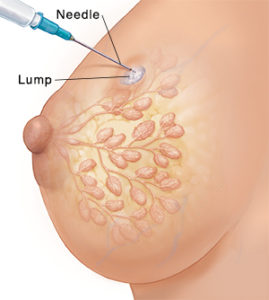
FNA is a way of taking a few cells from inside your breast using a syringe with a fine needle. This is done after the mammogram shows anything abnormal in your breasts.
- Before an FNAC, you will be asked to take all your clothes off your top half and you shall be asked to lie down.
- The nurse/doctor will gently push a needle into your breast and pull out some cells using the syringe. This needle is moved in and out multiple times to create the suction which collects the cells.
- The sample cells are collected on a slide and viewed under the microscope, which can establish the diagnosis of cancer in most cases.
- When the needle is taken out, a mesh (like a sticker) will be put over the place where the needle went in, which can be removed the next day.
Core Biopsy
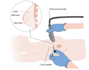 Core biopsy is a method of taking out a small part of the inside of your breast through a small cut in your skin. In this process, a ‘core biopsy gun’ is used. The doctor/nurse doing the core biopsy will decide which part to take out by looking at your mammogram.
- Before a core biopsy, an injection shall be used to numb the area before the cut is made.
- A needle will be inserted through the cut and a small part from inside your breast will be taken out. You may hear a loud clicking noise.
- After the biopsy, a plaster or other covering over the cut shall be pasted where the needle went in. This can be removed in 2-3 days.
- Once the numbing injection has worn off, the breast might hurt, for which you can take pain-killers. You might also see a bruise on your breast.
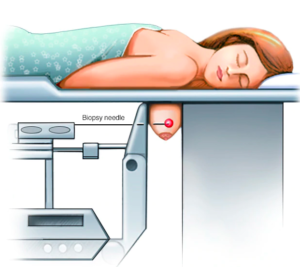
Please note that sometimes for core biopsy, more pictures are needed to get the needle to just the right place. For this, the mammogram machine is used. Your breast will be pressed in the mammogram plates while the core biopsy is taken. This can be a bit uncomfortable and painful and is called Stereotactic Core Biopsy.
THE TEST RESULTS
Staff at the hospital will let you know when and how they will tell you what your tests show. The wait time differs from clinic to clinic. Some also tell the result on the same day. You might want to have a close friend/family member to be by your side when the results arrive.
- If reports show no cancer: This means that the changes in the breasts are either normal or there is a Benign Condition. The doctor shall let you know if you need any treatment for a benign condition.
- If reports show the presence of cancer: From our conversations with women, we have come to understand the myriad of emotions women have undergone when they were first told they had breast cancer. While some got scared of the thought that they were going to die, some blacked out at the news, some felt angry and shocked and asked themselves- ‘Why me?’, some refused to believe it stating that ‘They have always felt fine and this can’t happen to them’, some took it optimistically and got relieved that cancer has been found and that it is going to be treated while some went completely numb. Every woman coped with the diagnosis of breast cancer in different ways and it is completely natural to feel hopeful on some days, and very low or anxious on others.
THE NEXT STEPS
The surgical oncologist will assess whether she/he shall perform the surgery first, or chemotherapy first. If she/he plans to operate first, they shall either go for breast-conserving surgery or mastectomy, depending on mammography findings. If she/he plans chemotherapy first, she/he will do a core biopsy, send the specimen for lab-testing and after confirming the diagnosis, start chemotherapy. We shall talk about ‘coping up with the news’, ‘informing family/children’, ‘undergoing treatment’ and more, in our next and upcoming series of articles.